Ras Family of Proteins in Plasmodium Falciparum Ookinete
- Inquiry
- Open Admission
- Published:
Plasmodium falciparum ookinete expression of plasmepsin Seven and plasmepsin X
Malaria Periodical volume fifteen, Article number:111 (2016) Cite this commodity
Abstract
Groundwork
Plasmodium invasion of the mosquito midgut is a population bottleneck in the parasite lifecycle. Interference with molecular mechanisms by which the ookinete invades the mosquito midgut is 1 potential arroyo to developing malaria transmission-blocking strategies. Plasmodium aspartic proteases are one such course of potential targets: plasmepsin IV (known to be present in the asexual stage food vacuole) was previously shown to be involved in Plasmodium gallinaceum infection of the mosquito midgut, and plasmepsins VII and plasmepsin X (not known to be nowadays in the asexual phase food vacuole) are upregulated in Plasmodium falciparum mosquito stages. These (and other) parasite-derived enzymes that play essential roles during ookinete midgut invasion are prime candidates for transmission-blocking vaccines.
Methods
Reverse transcriptase PCR (RT-PCR) was used to determine timing of P. falciparum plasmepsin Seven (PfPM Vii) and plasmepsin X (PfPM X) mRNA transcripts in parasite mosquito midgut stages. Poly peptide expression was confirmed by western immunoblot and immunofluorescence assays (IFA) using anti-peptide monoclonal antibodies (mAbs) against immunogenic regions of PfPM Vii and PfPM X. These antibodies were too used in standard membrane feeding assays (SMFA) to determine whether inhibition of these proteases would affect parasite transmission to mosquitoes. The Isle of mann–Whitney U test was used to analyse mosquito transmission assay results.
Results
RT-PCR, western immunoblot and immunofluorescence assay confirmed expression of PfPM Seven and PfPM X in mosquito stages. Whereas PfPM VII was expressed in zygotes and ookinetes, PfPM Ten was expressed in gametes, zygotes, and ookinetes. Antibodies against PfPM VII and PfPM X decreased P. falciparum invasion of the musquito midgut when used at high concentrations, indicating that these proteases play a office in Plasmodium mosquito midgut invasion. Failure to generate genetic knockouts of these genes limited determination of the precise role of these proteases in parasite transmission but suggests that they are essential during the intraerythrocytic life bicycle.
Conclusions
PfPM VII and PfPM 10 are present in the mosquito-infective stages of P. falciparum. Standard membrane feeding assays demonstrate that antibodies against these proteins reduce the infectivity of P. falciparum for mosquitoes, suggesting their viability as transmission-blocking vaccine candidates. Further report of the role of these plasmepsins in P. falciparum biology is warranted.
Groundwork
Despite reduced incidence and bloodshed rates malaria continues to have a significant global affect with more than 200 million people infected and a reported 438,000 deaths in 2015 alone [ane]. Malaria is initiated when sporozoites are injected into a human from the bite of a Plasmodium-infected anopheline mosquito, with severe disease almost often due to Plasmodium falciparum. Increasing resistance to common anti-malarial drugs and the lack of an effective vaccine heighten the importance of malaria every bit a global scourge requiring new approaches, including novel vaccine approaches to simultaneously target multiple parasite stages [two]. In improver to sporozoite-targeting malaria vaccine approaches that aim to prevent initial human infection, complementary lines of malaria vaccine research have also focused on so-called transmission-blocking vaccines or, as newly articulated "vaccines that interrupt malaria transmission" [2], which aim to preclude humans with Plasmodium gametocytaemia from infecting the musquito vector [3, 4]. Transmission-blocking vaccines target proteins expressed on or secreted by sexual stage parasites, which develop in the musquito midgut [five].
Anopheles species mosquitoes are the definitive hosts of Plasmodium parasites, which obligatorily complete development from sexual stages to sporozoites within the mosquito before manual to humans. Upon ingestion by the musquito, environmental changes encountered in the midgut stimulate mature gametocyte emergence from infected erythrocytes, in a process known equally gametogenesis [half-dozen, seven]. Sexually dimorphic gametes fuse to generate zygotes in the midgut lumen. Zygotes and then undergo sexual recombination and meiotic replication followed past transformation into polarized, motile ookinetes [8–x]. Ookinetes penetrate the midgut epithelium and form oocysts on the basal lamina. I ookinete that penetrates the midgut wall to form an oocyst has the potential to generate thousands of sporozoites, the form of the parasite that infects humans [11, 12]. Ookinete invasion of musquito midgut is an important process for malaria manual, but little is known nigh the molecular mechanisms involved.
Ookinetes produce stage-specific proteins important for subsequent midgut invasion, such as chitinase [thirteen, fourteen], circumsporozoite- and thrombospondin-related adhesive protein [TRAP]-related protein (CTRP) [xv, xvi], von Willebrand A domain-related protein (WARP) [17, 18], Plasmodium 25/28 zygote/ookinete surface proteins (P25/28) [nine, nineteen, 20], secreted ookinete adhesive protein (SOAP) [16, 21], and membrane assault perforin (MAOP) [22] amid others. Because proteases play of import roles during parasite infection of and development in the musquito, they were considered every bit potential transmission-blocking vaccine targets [23–28]. Transcriptomic information suggested that Plasmodium aspartic proteases, known as plasmepsins, are expressed in sexual stage parasites [29]. The P. falciparum genome encodes ten Plasmodium aspartic proteases known every bit plasmepsins [30]. Plasmepsins I, II, 4 and HAP are present in the asexual blood stage parasite food vacuole and are involved in haemoglobin degradation in the food vacuole of blood stage parasites [30–35]. In the endoplasmic reticulum of asexual claret stage parasites, Plasmepsin Five processes proteins and directs export of effector proteins [36]. Plasmepsin Six plays an as-however undefined merely of import role in parasite sporogonic development, particularly in early on oocyst development in Plasmodium berghei [37]. Plasmepsin IV, in add-on to its known role in haemoglobin degradation, is involved in Plasmodium ookinete invasion of the mosquito midgut [38].
Transcriptomic data accept shown that P. falciparum plasmepsin Seven (PfPM 7) mRNA is present in gametocytes, and plasmepsin X (PfPM Ten) mRNA is present during both in gametocytes and in zygotes and ookinetes [29]. The biological functions of PfPM VII and PfPMX remain unknown. Based on these observations, this study aimed to test the hypothesis that PfPM Seven and PfPM X are targets of blocking mosquito midgut infection by P. falciparum. Such data would support the notion that these proteins would contribute to the interaction of the P. falciparum ookinete with the anopheline midgut and provide the ground for further development of these molecules as components of a malaria transmission-blocking vaccine.
Methods
Parasites and mosquitoes
Plasmodium falciparum strain NF54 from a main cell depository financial institution was used in this study, kindly provided under a material transfer agreement with Sanaria, Inc, Rockville, Physician, Us. Parasites were maintained in asexual culture according to standard protocol [39]. Gametocytes and gametes were cultured in vitro co-ordinate to the Ifediba and Vanderberg [40] modification of the Trager and Jensen method [41]; zygotes and ookinetes were cultured and purified as described [42–44].
Anopheles gambiae and Anopheles stephensi used in this study were a generous gift from Dr. Anthony James (University of California Irvine, Irvine, CA USA). Mosquitoes were maintained in an enclosed insectary at 26 °C and fourscore % humidity with an automated 12-h light–nighttime cycle according to standard CDC protocol [45]. Mosquitoes used in this study were closely monitored and controlled according to the protocol for non-vertebrate animal subjects approved past the UCSD Institutional Animal Care and Employ Committee (IACUC).
DNA/RNA isolation and RT-PCR
Plasmodium parasites were either generated in vitro or isolated ex vivo from infected mosquitoes. For ex vivo-isolated mosquito-stage parasite samples, midguts from mosquitoes were dissected and homogenized; 5 midguts were pooled per sample at 24 h mail-engorgement. Genomic Deoxyribonucleic acid was isolated using NucleoSpin Blood (Macherey–Nagel, Bethlehem, PA, U.s.). Full RNA was isolated using RNeasy (Qiagen, Valencia, CA, United states of america) and contaminating DNA was removed using DNA-gratuitous (Ambion, Austin, TX, Us) according to manufacturer'southward instructions. Reverse transcription was completed using factor-specific primers for PfPM VII, PfPM X and Pfs25 with SuperScript 3 first-strand synthesis organization (Invitrogen, Carlsbad, CA, USA) according to manufacturer's instructions. PCR on resulting cDNA was done using Platinum PCR SuperMix Loftier Allegiance (Invitrogen, Carlsbad, CA, USA) with 250 nM of the aforementioned gene-specific primers for: Pfs25 (Fwd five′-tgcgaaagttaccgtggatactg-3′; Rev 5′-tgcgaaagttaccgtggatactg-iii′), PfPM VII (Fwd five′-gcgccatgggtaaaaatgaagaattcacgaatccttattcc-3′, Rev 5′-gcgctcgagccttaaggttacatttcttttacttctaac-3′) and PfPM 10 (Fwd 5′- gtgatgaagaaagttacgttatatttgacacagg-3′; Rev 5′-gctcttgctactccaaccatagaagg-3′). Thirty-v cycles were run with an annealing temperature of 55 °C and an extension temperature of 68 °C.
Production of recombinant PfPM 7 and PfPM 10 in Escherichia coli
PfPM VII and PfPM X genes were amplified from genomic NF54 Dna to generate a factor that lacked the signal peptide. PCR products were gel purified on a 0.8 % agarose gel using the PureLink gel extraction kit (Invitrogen, Carlsbad, CA, The states), ligated into pCR4-TOPO (Invitrogen, Carlsbad, CA, The states), transformed into Top10 competent cells (Invitrogen, Carlsbad, CA, USA) and sequence-verified (Eton Bioscience, San Diego, CA, United states of america). PfPM VII and PfPM X genes were and so cloned into expression vectors pET32 and pGEX 4T-1 (GE Healthcare, Piscataway, NJ, The states), respectively.
The recombinant PfPM Vii-HIS-tagged (rPfPM VII-HIS) and PfPM X-GST-tagged (rPfPM 10-GST) fusion proteins were expressed in Rosetta (Merck/Novagen, Darmstadt, Germany) competent cells according to standard protocol [46]. Briefly, competent cells were transformed with 250–500 ng of purified plasmid DNA, streaked on LB agar plates embedded with 100 µg/ml ampicillin and allowed to grow overnight at 37 °C. Fresh colonies were inoculated and grown to OD600 0.five–1. Protein expression was then induced with 0.3 mM isopropyl β-D-1-thiogalactopyranoside (IPTG) for 0.5–18 h at 18 °C.
For rPfPM Seven and rPfPM X poly peptide verification, inclusion bodies were purified using BugBuster extraction reagent (Merck/Novagen, Darmstadt, Deutschland) and run on a 4–20 % Tris–Glycine SDS-PAGE gel that was then stained with Coomassie blue. A stained SDS-Page gel slice consistent with the predicted size of rPfPM 7-HIS and rPfPM X-GST fusion protein was excised and processed for mass spectrometry analysis (The Scripps Research Institute, La Jolla, CA, Usa). The gel slices were destained and proteins were reduced with 10 mM DTT, alkylated with 55 mM iodoacetamide and digested in-gel with trypsin as previously described [47, 48]. Samples were analysed by mass spectrometry, and identified peptides were searched by Smash confronting the P. falciparum genome.
Product of monoclonal antibodies confronting PfPM Vii and PfPM X
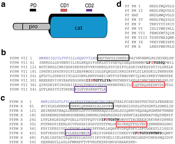
PfPM Seven peptide sequences EEFTNPYSIRKKDI, IQPDEQSEEDNVDG, and KIGFVRSKRNVTLR were derived from predicted immunogenic regions of the PfPM VII protein sequence using resources freely available from the Immune Epitope Database (iedb.org). PfPM X peptide sequences KKLQKHHESLKLGDVKYYV, VRNQTFGLVESES, and LKESYWEVKLD were derived from predicted immunogenic regions of the PfPM 10 poly peptide sequence (Fig. 1). These synthetic peptides were created with an N-terminal cysteine to facilitate coupling to bovine serum albumin (BSA) equally the antigenic carrier protein. Peptide composition was confirmed by mass spectrometry, and HPLC-purified peptides were used to immunize mice. The resulting hybridoma supernatants were screened against three sets of BSA-conjugated synthetic peptides, the aforementioned used to immunize mice, by enzyme linked immunosorbent analysis (ELISA; A&G Pharmaceuticals, Columbia, MD, USA). Secondary screening of ELISA-positive supernatant was performed past western immunoblot assay against total-length rPfPM VII-HIS or rPfPM X-GST. Hybridoma lines generating mAb that screened positive in both ELISA and Western immunoblot assays were grown in Dulbecco's modified eagle medium (DMEM, CellGro, Herndon, VA, USA) supplemented with 10 % fetal calf serum. Antibodies were either concentrated from hybridoma supernatant or purified mAb in PBS (A&G Pharmaceuticals, Columbia, MD, USA).

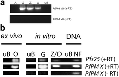
Plasmepsin Seven and Plasmepsin X mRNA was detected in Plasmodium falciparum sexual stage parasites. a Total RNA isolated from in vitro-cultivated asexual stages (A), gametocytes (G), zygotes (Z), ookinetes (O), and uninfected human erythrocytes (uB). Samples were contrary transcribed and amplified using primers specific for PfPM VII (+RT). Samples that were not reverse transcribed (−RT) and amplified with PfPM Vii-specific primers did not generate PCR product. b Full RNA isolated ex vivo from ookinete-containing mosquito midguts (O) or uninfected human blood (uB), in vitro-cultivated gametocytes (G) and mixed zygotes and ookinetes (Z/O), equally well as Deoxyribonucleic acid from P. falciparum (NF) was isolated. Samples were contrary transcribed and amplified using primers specific for PfPM 10 and pfs25 (+RT). Samples that were not reverse transcribed (−RT) and amplified with PfPM X-specific primers did not generate PCR product
After screening against rPfPM Seven, 2 lines, 1B4 and 2B1, generated antibodies directed against each catalytic domain of PfPM VII. After screening against rPfPM X, one line, 6A9, generated antibody directed confronting the pro-enzyme domain, and one line, 3G6, generated antibiotic directed against the catalytic domain (Fig. 2). mAb 6A9 was directed against a peptide region unique to PfPM X while 3G6 is directed against a peptide region that is moderately conserved with PfPM IX (Fig. two).

Peptide monoclonal antibodies directed against Plasmodium falciparum Plasmepsin 7 and Plasmepsin X. a Schematic representation of Plasmepsin showing the predicted indicate peptide (blank), pro-enzyme domains (pro), and catalytic domain (cat). Peptide monoclonal antibodies designed against three regions of PfPM Seven and PfPM X are designated prodomain (PD, black), catalytic domain 1 (CD1, red) and catalytic domain 2 (CD2, royal). b Protein sequence of PfPM Vii and c. PfPM X showing the predicted indicate peptide (blueish) and the predicted pro-enzyme domain (italics); the ii active aspartic acid residues (red) are constitute within conserved regions (bold). Monoclonal antibody targets to prodomain (black), CD1 (red), and CD ii (purple). d Alignment of 3G6 peptide target between all P. falciparum plasmepsins shows moderate conservation between PfPM X and PfPM 9 at this site
Immunofluorescence analysis
Stock-still, permeabilized P. falciparum gametocytes, gametes, zygotes and ookinetes were analysed by IFA. Parasites were fixed on drinking glass slides with 100 % acetone at −20 °C for at to the lowest degree 20 min and then rehydrated by two changes of PBS for 5 min each at room temperature. For membrane permeabilization and blocking of nonspecific bounden, fixed cells were incubated in PBS supplemented with iii % bovine serum albumin and 0.1 % Triton X-100 for ane h at room temperature. The preparations were then incubated with mAb (ane:1000 dilution) for 1 h at room temperature followed by either FITC-conjugated anti-mouse IgG or Alexa Fluor 488 rabbit anti-mouse IgG (1:200 dilution) (Molecular Probes, Invitrogen, USA). Nuclei were visualized with 300 nM DAPI (4, 6-diamidino-two-phenylindole) (Pierce Biotechnology, Rockford, IL, The states). Slides were washed an additional six times in Tris-buffered saline (TBS) for a total of 30 min so mounted with coverslips using Dako mounting medium (Dako, Carpinteria, USA). Preparations were examined past deconvolution microscopy using an Olympus BX51 fluorescence microscope and Olympus DP71 camera (Olympus, Center Valley, CA, Usa).
Immunoelectron microscopy localization of PfPM VII and PfPM X
Cells were fixed with two.v % glutaraldehyde in 0.1 K cacodylate buffer for 2 h at room temperature, postfixed in i % OsO4 in 0.one G cacodylate buffer (1 h) at room temperature, and embedded in Threescore-112 (Ladd Enquiry, Williston, VT), as described previously [49]. Cryosections were made, applied to grids, blocked in 1 % BSA in PBS for 1 h, incubated in 1:500 dilution of mAb confronting PfPM Seven, PfPM X or IgG isotype control, done and incubated with anti-mouse IgG conjugated to five nm colloidal gold particles as described previously [50]. Stained sections were examined using a Philips CM-x electron microscope.
Western immunoblot analysis
Plasmodium falciparum parasites were purified, pelleted and resuspended in 250 µl of lysis buffer (4 M urea, 0.4 % Triton Ten-100, l mM Tris, 5 mM EDTA, x mM MgSO4, pH 8.0) supplemented with Complete protease inhibitor cocktail (Roche Applied Sciences, Indianapolis, IN, United states of america). Parasites were processed by 3 cycles of freeze–thaw lysis followed by sonication on water ice for 5 min in xxx s bursts using a Misonix Sonicator 3000 with an output setting of 7 (Misonix, Farmingdale, NY, Usa). Poly peptide concentrations were determined by BCA assay (Bio-Rad, Hercules, CA, U.s.a.). 100 µg of each sample was mixed with Laemmli SDS-loading buffer (160 mM Tris, 10 % SDS, 20 % glycerol, v % two-mercaptoethanol, 0.01 % bromophenol blue, pH 8.0) and boiled for ten min. Proteins were separated on Novex ten–20 % SDS-PAGE mini-gels (Invitrogen, Carlsbad, CA 92008 USA) and transferred to nitrocellulose membranes. Membranes were blocked in TBS/five % non-fat milk/0.05 % Tween-twenty, pH eight.0 for 1 h. Blots were probed with primary antibody diluted 1:2000 in blocking buffer for ane h. Following six 10-min washes in blocking buffer, blots were probed with peroxidase-conjugated anti-mouse secondary antibody diluted one:20,000 in blocking buffer. Blots were washed half dozen times in blocking buffer, twice in TBS and then developed with chemiluminescent substrate (KPL, Gaithersburg, MD, Usa).
Membrane feeding assays
Standard membrane feeding assays (SMFA) were performed to make up one's mind the ability of antibodies confronting Pf PMVII and PfPMX to impact infectivity of P. falciparum for mosquitoes. One twenty-four hour period prior to the assay, female An. stephensi or An. gambiae mosquitoes aged iii–7 days post-emergence were segregated into cartons of 40–60 mosquitoes, and starved overnight. Mature P. falciparum gametocytes were examined for their ability to exflagellate and only those cultures with at least 10 exflagellating centres per 40X field were used for SMFA. Cultures with mature gametocytes were mixed with fresh human serum and red blood cells, plus antibodies, then fed to An. stephensi or An. gambiae using h2o-jacketed glass membrane feeders every bit previously described [14]. Isotype immunoglobulin G2b (IgG2b) was used every bit a negative control. Twenty minutes afterward the start of the feed, membrane feeders were disengaged and not-engorged mosquitoes were removed from cartons. P. falciparum-infected mosquitoes were kept in a secured incubator dissever from non-infected mosquitoes. Infected mosquitoes were fed with 8 % fructose/0.05 % p-aminobenzoic acid in sterile water ad libitum and maintained at 26–28 °C and eighty % relative humidity [51, 52].
On day 8–ten mail claret meal, mosquito midguts were dissected, stained with mercurochrome and examined with a light microscope for the presence of oocysts [52]. All manipulations were done in accordance with UCSD IACUC-approved protocol for non-vertebrate research animals. Differences in infection rate and geometric means between test mAb and negative control groups were assessed using the not-parametric Isle of man–Whitney U test. Samples were considered to be statistically meaning at a p value ≤0.01.
Results
PfPM Seven and PfPM 10 mRNA was transcribed in P. falciparum sexual stages
RT-PCR of RNA isolated from P. falciparum sexual stage parasites generated in vitro and dissected from mosquitoes ex vivo demonstrated that PfPM Vii and PfPM X mRNA was detected in gametocytes and mixed zygotes/ookinetes (Fig. i). Conventional PCR of RNA samples not treated with reverse transcriptase did not produce PCR product and demonstrated that RNA samples were Dna free (Fig. ane).
PfPM VII and PfPM X protein expression in sexual stage parasites
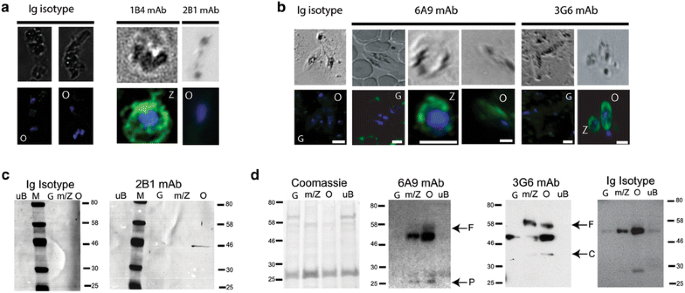
IFA of P. falciparum sexual stage parasites using mAbs directed against PfPM 7 and PfPM X demonstrated diffuse, cytoplasmic localization in P. falciparum zygotes and ookinetes merely not gametocytes (Fig. three). Neither PfPM 7 nor PfPM 10 were found to exist localized within specific sub-cellular compartments, such as micronemes or endoplasmic reticulum, or the ookinete prison cell surface as determined by immunoelectron microscopy.

PfPM VII and PfPM X expression in Plasmodium falciparum sexual stage parasites demonstrated by IFA and western immunoblot. IFA of in vitro-cultivated parasites was performted using main antibodies 1B4, 2B1, 6A9, 3G6 or isotype IgG2b and subsequently labelled with Alexa Fluor 488-labelled anti-mouse antibodies (greenish); nuclei are visualized with DAPI (blue). a IFA of in vitro-cultivated P. falciparum demonstrated PfPM 7 expression in zygotes (Z) and ookinetes (O). b IFA of in vitro-cultivated P. falciparum demonstrated PfPM Ten expression in zygotes (Z) and ookinetes (O) but not gametocytes (One thousand). c Western immunoblot analysis of total poly peptide isolated from P. falciparum sexual phase parasites. Antibodies directed confronting PfPM VII recognized a ~46 kDa protein expressed in ookinetes (O) but non gametocytes (1000). This protein is between the predicted sizes of full length PfPM 7 at 52 kDa and the catalytic domain at 43 kDa. d Western immunoblot analysis of full poly peptide isolated from P. falciparum sexual stage parasites. Both 6A9 and 3G6 recognized protein expressed in mixed gametes and zygotes (yard/Z) and ookinetes (O) but not gametocytes (One thousand). Bands are consistent with the predicted sizes of full length PfPM X at 61 kDa (←F), the PfPM Ten catalytic domain at 36 kDa (←C), and the PfPM 10 proenzyme domain at 23 kDa (←P). IgG isotype control did non recognize the 61 kDa or 23 kDa bands on western immunoblot but continued to recognize the bands at 42–55 kDa and 27–30 kDa
Western immunoblot confirmed IFA results demonstrating PfPM VII and PfPM X protein expression in sexual stage parasites. Antibody 2B1 recognized a single ~ 46 kDa band in ookinetes simply no other sexual stage parasite; this protein is close to the predicted 52 kDa size of the total length protein and the 42 kDa size of the PfPM 7 catalytic domain (Fig. 3). Antibodies 6A9 and 3G6 recognized multiple bands in mixed gamete/zygote samples and ookinete samples, simply not gametocyte samples. Antibiotic 6A9 recognized ii bands: a 56–60 kDa protein, consistent with the predicted 61 kDa size of full length PfPM X, and a faint 17–25 kDa protein, consistent with the predicted 23 kDa size of the PfPM X pro-enzyme domain. Antibody 3G6 as well recognized a 56–lx kDa protein in addition to a faint 32–40 kDa poly peptide, consistent with the predicted 36 kDa size of the catalytic domain. Of annotation, 6A9 and 3G6 recognized two bands in parasite lysate, one at 42–55 kDa and the second at 27–xxx kDa; these bands were besides recognized by IgG isotype control antibody, indicating that these protein bands are non-specific (Fig. 3).
Antibodies directed against PfPM VII or PfPM X moderately decreased Plasmodium falciparum manual to Anopheles in SMFAs
To decide whether antibodies against either PfPM VII or PfPM X could affect P. falciparum transmission to mosquitoes, female person An. stephensi and An. gambiae were fed infectious gametocytes mixed with 1B4, 2B1, 6A9, 3G6 or isotype IgG negative control antibodies, respectively, in SMFAs (Table ane, 2). Mosquito midguts were dissected 8–x days post-blood repast to determine prevalence and intensity of infection.
The presence of antibodies directed against either PfPM Seven or PfPM X in an infectious blood repast significantly decreased P. falciparum transmission to mosquitoes at high antibody concentrations (Tables 1, two). Mosquitoes fed an infectious bloodmeal with 200 µg/ml of 1B4 or 400 µg/ml of 2B1 had a 35–71.four or 9.7–23.5 % reduction in prevalence compared to groups fed with IgG isotype control antibody (Tabular array one). Additionally, oocyst intensity of infected mosquitoes fed with1B4 or 2B1 was reduced by 81–97 or 56–65 % compared to IgG isotype control (p value < 0.01) (Table ane). Mosquitoes fed an infectious blood repast with 100 or 200 µg/ml of 3G6 had a 18 or 33 % reduction in prevalence compared to IgG isotype control (Tabular array 2). Additionally, the oocyst intensity of infected mosquitoes fed with 100 μg/ml of 3G6 was reduced by 35 % compared to IgG isotype control, the oocyst intensity of infected mosquitoes fed with 200 μg/ml of 3G6 was reduced by 42 % compared to IgG isotype controls (p value < 0.01) (Table 2). The presence of 200 µg/ml of 6A9 in an infectious bloodmeal only reduced P. falciparum transmission to mosquitoes by thirteen %, and oocyst intensity was comparable to IgG isotype controls (Table 2).
Further attempts were made to elucidate the precise role of PfPM VII and PfPM X during P. falciparum sexual development and transmission to mosquitoes. Unfortunately, we were unable to produce active, recombinant protease in either an Due east. coli-based expression organization or a cell-free wheat germ expression system. The generation of PfPM Seven and PfPM Ten knockout parasites were unsuccessful (Additional files 1, 2).
Give-and-take
The data reported hither signal that that PfPM VII and PfPM X are expressed in ookinetes and contribute to P. falciparum transmission to Anopheles mosquitoes. This is the first observation that these plasmepsins play an important role in Plasmodium infection of mosquitoes. Transcriptomic data from all the life cycle stages of P. falciparum demonstrate mRNA expression of PfPM VII and PfPM X in sexual phase forms. These previously published microarray information were confirmed by RT-PCR on in vitro-cultivated gametocytes, gametes, zygotes and ookinetes and ex vivo-harvested zygotes and ookinetes. Anti-peptide monoclonal antibodies directed confronting PfPM Seven or PfPM Ten demonstated that these proteins are expressed in sexual phase parasite forms. Interestingly, PfPM 7 and PfPM X mRNAs are expressed in P. falciparum gametocytes, only protein was not detected until zygote and gamete development, respectively. This inconsistency between mRNA and poly peptide expression was not explored in this work, but this design suggests that PfPM Seven and PfPM Ten protein expression may be regulated by translational repression, a mechanism used to regulate other Plasmodium sexual stage proteins [53–58]. Similar observations accept been seen with Pfs25 and chitinase, whose mRNAs but not proteins take been detected in gametocytes [fifty, 59].
Another mechanism to regulate protease activity is zymogen processing. Plasmepsins are known to harbour a proenzyme domain that functions to maintain the enzyme in a catalytically inactive country. Plasmepsins only become catalytically active when the proenzyme domain is cleaved and separated from the catalytic domain. Both PfPM 7 and PfPM X contain predicted proenzyme domains. Western immunoblot of all three mAbs targeting PfPM VII just detected one poly peptide in parasite lysate; this finding was unexpected as PfPM VII also contains a predicted proenzyme domain. Additionally, the poly peptide recognized is smaller than the predicted size of the full-length enzyme only large than the predicted size of the PfPM 7 catalytic domain. Although we would wait to encounter two bands corresponding to the full-length protein and the proenzyme domain alone, it is possible that PfPM VII is not candy and activated in the ookinete. If PfPM Seven is secreted or prison cell-surface associated, a hypothesis non currently supported by IFA or immunoelectron data, it is possible that proenzyme domain is cleaved outside of the parasite. Given this event, it is likely that the total-length protein is processed at a site upstream of the mAb target region just that the proenzyme domain has not nevertheless been cleaved in the ookinete, resulting in a smaller-than expected full-length protein. Western immunoblot analysis demonstrated that 6A9, which targets the PfPM X proenzyme domain, recognized proteins consequent with the sizes of the full-length enzyme and the cleaved pro-enzyme domain while 3G6, which targets the catalytic domain, recognized proteins consistent with the sizes of the full-length enzyme and the cleaved catalytic domain. This observation suggests that PfPM X is processed, and potentially catalytically agile, during P. falciparum sexual stage development. Western immunoblot demonstrate that both PfPM Vii and PfPM X are candy, and that PfPM X is likely catalytically active in the ookinete.
The majority of proteins that are both important for Plasmodium manual to mosquitoes and good targets for transmission-blocking vaccines are either localized to the parasite cell surface or secreted from the ookinete. Cell-surface proteins, such as P25/P28, mediate parasite-host interactions and may be essential components of signalling-pathways important for ookinete motility [15, nineteen, 21, 60]. Similarly, ookinete-secreted proteins and enzymes, such as chitinase, CTRP and WARP, modify the musquito midgut environs to allow Plasmodium invasion and infection of the mosquito midgut [xiii, fourteen, 17, xviii]. Neither PfPM VII nor PfPM X localized to the parasite cell surface or micronemes. IFA and immunoelectron microscopy demonstrated diffuse cytoplasmic localization of mAbs directed confronting PfPM 7 and PfPM X.
Previous studies showed that pepstatin A, an aspartic protease-specific inhibitor, ALLN and ALLM, two plasmepsin convertase inhibitors, interrupted Plasmodium transmission to mosquitoes [38]. The presence of antibodies directed against PfPM VII or PfPM X significantly reduced P. falciparum infection of Anopheles. However, relatively high concentrations of 1B4, 2B1 and 3G6 were required to achieve an issue. Information technology is possible that the antibodies had low affinity, resulting in the depression level of interruption of the transmission. It is too possible that these Plasmepsins were important, but not essential, for P. falciparum transmission to Anopheles mosquitoes, consequent with previous reports with a P. berghei Plasmepsin Vii knockout mutant [61]. Every bit, if non more likely is the possibility that loss of PfPM Vii or PfPM X function in the presence of mAb was compensated for by the activeness of related proteases, such as Plasmepsin IX, which are similarly upregulated in sexual stage parasite forms [29]. The equivocal result seen in SMFAs using 6A9, directed against the PfPM X proenzyme domain, is expected if the proenzyme domain of PfPM X is broken and no longer associated with the activated catalytic domain in ookinetes. These information point that there is yet much to learn virtually the office of plasmepsins in Plasmodium transmission to mosquitoes. Further work is needed to explore these interesting possibilities.
Conclusions
Knowledge of the Plasmodium sexual bike and the molecular mechanisms underlying ookinete invasion of the mosquito midgut is essential for understanding parasite developmental programmes and parasite-musquito interactions. In this study, we accept investigated the expression and functional significance of PfPM VII and PfPM Ten in malaria transmission to mosquitoes. Nosotros demonstrated that these proteins are expressed in P. falciparum ookinetes and that antibodies directed against these proteins reduced parasite manual to Anopheles mosquitoes when used at high concentration in mosquito infection experiments. This observation suggests that PfPM VII and PfPM Ten facilitate Plasmodium infection of mosquitoes. This information complements discoveries demonstrating that P. gallinaceum plasmepsin 4 was important for parasite infection of the musquito vector [38] and P. berghei plasmepsin VII was dispensable in its life cycle [61]. Together, these results suggest that plasmepsins expressed in the Plasmodium ookinete function in means singled-out from plasmepsins expressed in asexual parasites. These findings raise a number of interesting questions. What other plasmepsins are expressed in sexual stage parasites? If these plasmepsins are involved in midgut invasion or sexual stage parasite development, what pathways are involved? Further investigation of the role(s) of plasmepsins during sexual evolution and invasion of mosquitoes will probable lead to novel insights into Plasmodium biological science and provide new targets for manual-blocking vaccines.
Abbreviations
- PfPM VII:
-
Plasmodium falciparum plasmepsin Vii
- PfPM Ten:
-
Plasmodium falciparum plasmepsin 10
- RT-PCR:
-
reverse transcriptase polymerase chain reaction
- SMFA:
-
standard membrane feeding assay
- Pfs25:
-
Plasmodium falciparum 25 kDa surface molecule
- IFA:
-
immunofluorescence analysis
- ELISA:
-
enzyme-linked immunosorbent assay
- mAb:
-
monoclonal antibody
References
-
WHO: World Malaria Report 2015. Geneva, Globe Wellness Organization; 2015.
-
malERA Consultative Group on Vaccines. A research agenda for malaria eradication: vaccines. PLoS Med. 2011;8:e1000398.
-
Nunes JK, Woods C, Carter T, Raphael T, Morin MJ, Diallo D, et al. Development of a manual-blocking malaria vaccine: progress, challenges, and the path forward. Vaccine. 2014;32:5531–9.
-
DeWeerdt Due south. Vaccines: the take-home lesson. Nature. 2012;484:S24–v.
-
Kaslow DC. Manual-blocking vaccines. Chem Immunol. 2002;lxxx:287–307.
-
Sinden RE, Canning EU, Bray RS, Smalley ME. Gametocyte and gamete development in Plasmodium falciparum. Proc R Soc Lond B. 1978;201:375–99.
-
Sinden RE, Hartley RH, Rex NJ. Gametogenesis in Plasmodium; the inhibitory effects of anticytoskeletal agents. Int J Parasitol. 1985;15:211–7.
-
Sinden RE, Billingsley PF. Plasmodium invasion of mosquito cells: militarist or dove? Trends Parasitol. 2001;17:209–12.
-
Billy LA, Ranford-Cartwright LC. Spreading the seeds of million-murdering death: metamorphoses of malaria in the musquito. Trends Parasitol. 2005;21:573–80.
-
Siden-Kiamos I, Louis C. Interactions betwixt malaria parasites and their mosquito hosts in the midgut. Insect Biochem Mol Biol. 2004;34:679–85.
-
Poudel SS, Newman RA, Vaughan JA. Rodent Plasmodium: population dynamics of early sporogony within Anopheles stephensi mosquitoes. J Parasitol. 2008;94:999–1008.
-
Sinden RE, Billingsley PF. Plasmodium invasion of mosquito cells: hawk or dove? Trends Parasitol. 2001;17:209–11.
-
Vinetz JM, Valenzuela JG, Specht CA, Aravind L, Langer RC, Ribeiro JM, et al. Chitinases of the avian malaria parasite Plasmodium gallinaceum, a form of enzymes necessary for parasite invasion of the mosquito midgut. J Biol Chem. 2000;275:10331–41.
-
Li F, Patra KP, Vinetz JM. An anti-chitinase malaria transmission-blocking single-chain antibody as an effector molecule for creating a Plasmodium falciparum-refractory mosquito. J Infect Dis. 2005;192:878–87.
-
Dessens JT, Beetsma AL, Dimopoulos G, Wengelnik Thou, Crisanti A, Kafatos FC, et al. CTRP is essential for mosquito infection past malaria ookinetes. EMBO J. 1999;eighteen:6221–7.
-
Nacer A, Underhill A, Hurd H. The microneme proteins CTRP and SOAP are not essential for Plasmodium berghei ookinete to oocyst transformation in vitro in a cell free system. Malar J. 2008;seven:82.
-
Yuda Grand, Yano One thousand, Tsuboi T, Torii M, Chinzei Y. von Willebrand Factor A domain-related protein, a novel microneme protein of the malaria ookinete highly conserved throughout Plasmodium parasites. Mol Biochem Parasitol. 2001;116:65–72.
-
Li F, Templeton TJ, Popov V, Comer JE, Tsuboi T, Torii M, Vinetz JM. Plasmodium ookinete-secreted proteins secreted through a mutual micronemal pathway are targets of blocking malaria transmission. J Biol Chem. 2004;279:26635–44.
-
Duffy PE, Kaslow DC. A novel malaria poly peptide, Pfs28, and Pfs25 are genetically linked and synergistic equally falciparum malaria transmission-blocking vaccines. Infect Immun. 1997;65:1109–xiii.
-
Gregory JA, Li F, Tomosada LM, Cox CJ, Topol AB, Vinetz JM, et al. Algae-produced Pfs25 elicits antibodies that inhibit malaria transmission. PLoS One. 2012;7:e37179.
-
Dessens JT, Siden-Kiamos I, Mendoza J, Mahairaki Five, Khater Due east, Vlachou D, Xu XJ, et al. SOAP, a novel malaria ookinete poly peptide involved in mosquito midgut invasion and oocyst development. Mol Microbiol. 2003;49:319–29.
-
Kadota K, Ishino T, Matsuyama T, Chinzei Y, Yuda G. Essential function of membrane-attack poly peptide in malarial transmission to mosquito host. Proc Natl Acad Sci. 2004;101:16310–5.
-
Huber Thou, Cabib E, Miller LH. Malaria parasite chitinase and penetration of the mosquito peritrophic membrane. Proc Natl Acad Sci. 1991;88:2807–10.
-
Sieber KP, Huber M, Kaslow D, Banks SM, Torii M, Aikawa M, et al. The peritrophic membrane as a bulwark: its penetration by Plasmodium gallinaceum and the outcome of a monoclonal antibody to ookinetes. Exp Parasitol. 1991;72:145–56.
-
Han YS, Thompson J, Kafatos FC, Barillas-Mury C. Molecular interactions between Anopheles stephensi midgut cells and Plasmodium berghei: the time bomb theory of ookinete invasion of mosquitoes. EMBO J. 2000;nineteen:6030–40.
-
Eksi South, Czesny B, Greenbaum DC, Bogyo Grand, Williamson KC. Targeted disruption of Plasmodium falciparum cysteine protease, falcipain 1, reduces oocyst product, not erythrocytic stage growth. Mol Microbiol. 2004;53:243–50.
-
Aly AS, Matuschewski K. A malarial cysteine protease is necessary for Plasmodium sporozoite egress from oocysts. J Exp Med. 2005;202:225–30.
-
Blackman MJ. Malarial proteases and host jail cell egress: an 'emerging' cascade. Prison cell Microbiol. 2008;10:1925–34.
-
Zhou Y, Ramachandran 5, Kumar KA, Westenberger S, Refour P, Zhou B. Bear witness-based note of the malaria parasite's genome using comparative expression profiling. PLoS Ane. 2008;3:e1570.
-
Coombs GH, Goldberg DE, Klemba 1000, Berry C, Kay J, Mottram JC. Aspartic proteases of Plasmodium falciparum and other parasitic protozoa as drug targets. Trends Parasitol. 2001;17:532–7.
-
Francis SE, Gluzman IY, Oksman A, Knickerbocker A, Mueller R, Bryant ML, et al. Molecular characterization and inhibition of a Plasmodium falciparum aspartic hemoglobinase. EMBO J. 1994;xiii:306–17.
-
Gluzman IY, Francis SE, Oksman A, Smith CE, Duffin KL, Goldberg DE. Order and specificity of the Plasmodium falciparum hemoglobin degradation pathway. J Clin Invest. 1994;93:1602–8.
-
Wyatt DM, Berry C. Activity and inhibition of plasmepsin IV, a new aspartic proteinase from the malaria parasite Plasmodium falciparum. FEBS Lett. 2002;513:159–62.
-
Banerjee R, Liu J, Beatty Westward, Pelosof Fifty, Klemba G, Goldberg DE. Iv plasmepsins are agile in the Plasmodium falciparum food vacuole, including a protease with an agile-site histidine. Proc Natl Acad Sci. 2002;99:990–five.
-
Le Bonniec S, Deregnaucourt C, Redeker V, Banerjee R, Grellier P, Goldberg DE, et al. Plasmepsin II, an acidic hemoglobinase from the Plasmodium falciparum nutrient vacuole, is active at neutral pH on the host erythrocyte membrane skeleton. J Biol Chem. 1999;274:14218–23.
-
Klemba M, Goldberg DE. Characterization of plasmepsin V, a membrane-bound aspartic protease homolog in the endoplasmic reticulum of Plasmodium falciparum. Mol Biochem Parasitol. 2005;143:183–91.
-
Ecker A, Bushell ES, Tewari R, Sinden RE. Reverse genetics screen identifies six proteins important for malaria development in the mosquito. Mol Microbiol. 2008;70:209–20.
-
Li F, Patra KP, Yowell CA, Matriarch JB, Chin K, Vinetz JM. Apical surface expression of aspartic protease plasmepsin 4, a potential transmission-blocking target of the Plasmodium ookinete. J Biol Chem. 2010;285:8076–83.
-
Read G, Hyde JE. Simple in vitro cultivation of the malaria parasite Plasmodium falciparum (erythrocytic stages) suitable for big-calibration preparations. Methods Mol Biol. 1993;21:43–55.
-
Ifediba T, Vanderberg JP. Complete in vitro maturation of Plasmodium falciparum gametocytes. Nature. 1981;294:364–vi.
-
Trager W, Jensen JB. Human malaria parasites in continuous civilization. Science. 1976;193:673–5.
-
Bounkeua V, Li F, Vinetz JM. In vitro generation of Plasmodium falciparum ookinetes. Am J Trop Med Hyg. 2010;83:1187–94.
-
Dinglasan RR, Alaganan A, Ghosh AK, Saito A, van Kuppevelt TH, Jacobs-Lorena Grand. Plasmodium falciparum ookinetes require mosquito midgut chondroitin sulfate proteoglycans for prison cell invasion. Proc Natl Acad Sci U S A. 2007;104:15882–seven.
-
Ghosh AK, Dinglasan RR, Ikadai H, Jacobs-Lorena M. An improved method for the in vitro differentiation of Plasmodium falciparum gametocytes into ookinetes. Malar J. 2010;9:194.
-
Gerberg EJ, Barnard DR, Ward RA, American Musquito Control Association. Transmission for mosquito rearing and experimental techniques. Rev. edn. Lake Charles, La.: American Musquito Control Association; 1994.
-
LaVallie ER, McCoy JM. Gene fusion expression systems in Escherichia coli. Curr Opin Biotechnol. 1995;six:501–6.
-
Hellman U, Wernstedt C, Gonez J, Heldin CH. Improvement of an "In-Gel" digestion procedure for the micropreparation of internal protein fragments for amino acid sequencing. Anal Biochem. 1995;224:451–v.
-
Rosenfeld J, Capdevielle J, Guillemot JC, Ferrara P. In-gel digestion of proteins for internal sequence analysis after 1- or ii-dimensional gel electrophoresis. Anal Biochem. 1992;203:173–9.
-
De Vries 50, Elenko E, McCaffery JM, Fischer T, Hubler L, McQuistan T, et al. RGS-GAIP, a GTPase-activating poly peptide for Galphai heterotrimeric Thousand proteins, is located on clathrin-coated vesicles. Mol Biol Cell. 1998;9:1123–34.
-
Vinetz JM, Dave SK, Specht CA, Brameld KA, Xu B, Hayward R, et al. The chitinase PfCHT1 from the human malaria parasite Plasmodium falciparum lacks proenzyme and chitin-binding domains and displays unique substrate preferences. Proc Natl Acad Sci. 1999;96:14061–6.
-
Carter R, Ranford-Cartwright L, Alano P. The civilisation and preparation of gametocytes of Plasmodium falciparum for immunochemical, molecular, and mosquito infectivity studies. Methods Mol Biol. 1993;21:67–88.
-
Sinden RE. The biology of Plasmodium in the mosquito. Experientia. 1984;twoscore:1330–43.
-
Mair GR, Lasonder East, Garver LS, Franke-Fayard BM, Carret CK, Wiegant JC, et al. Universal features of postal service-transcriptional cistron regulation are critical for Plasmodium zygote development. PLoS Pathog. 2010;6:e1000767.
-
Braks JA, Mair GR, Franke-Fayard B, Janse CJ, Waters AP. A conserved U-rich RNA region implicated in regulation of translation in Plasmodium female gametocytes. Nucleic Acids Res. 2008;36:1176–86.
-
Kooij TW, Matuschewski K. Triggers and tricks of Plasmodium sexual development. Curr Opin Microbiol. 2007;10:547–53.
-
Mair GR, Braks JA, Garver LS, Wiegant JC, Hall N, Dirks RW, et al. Regulation of sexual evolution of Plasmodium by translational repression. Scientific discipline. 2006;313:667–9.
-
del Carmen Rodriguez Thou, Gerold P, Dessens J, Kurtenbach K, Schwartz RT, Sinden RE, et al. Characterisation and expression of pbs25, a sexual and sporogonic phase specific poly peptide of Plasmodium berghei. Mol Biochem Parasitol. 2000;110:147–59.
-
Paton MG, Barker GC, Matsuoka H, Ramesar J, Janse CJ, Waters AP, et al. Construction and expression of a post-transcriptionally regulated malaria gene encoding a surface poly peptide from the sexual stages of Plasmodium berghei. Mol Biochem Parasitol. 1993;59:263–75.
-
Pradel G, Hayton K, Aravind L, Iyer LM, Abrahamsen MS, Bonawitz A, et al. A multidomain adhesion protein family expressed in Plasmodium falciparum is essential for transmission to the mosquito. J Exp Med. 2004;199:1533–44.
-
Siden-Kiamos I, Vlachou D, Margos 1000, Beetsma A, Waters AP, Sinden RE, et al. Distinct roles for pbs21 and pbs25 in the in vitro ookinete to oocyst transformation of Plasmodium berghei. J Cell Sci. 2000;113(Pt 19):3419–26.
-
Mastan BS, Kumari A, Gupta D, Mishra South, Kumar KA. Gene disruption reveals a dispensable office for plasmepsin Seven in the Plasmodium berghei life cycle. Mol Biochem Parasitol. 2014;195:10–3.
Authors' contributions
FL and VB designed and carried out molecular studies, mosquito feeding assays and statistical analysis, and drafted the manuscript. KP carried out recombinant protein activity assays and helped typhoon the manuscript. JMV conceived of the written report, and directed its design, coordinated the work and finalized the manuscript. All authors have read and approved the final manuscript.
Acknowledgements
The authors thank SL Hoffman for this NF54 strain of P. falciparum, AA James for providing A. gambiae mosquitoes. We thank T Tsuboi for producing rPfPM VII and rPfPM X using the cell-free wheat germ expression arrangement, MG Farquhar and G Kudlicka, Cadre Electron Microscopy Facility (funded by the US Public Health Service grants listed). We thank DA Fidock for advice with genetic knockout engineering science. We thank A. Lubar for figure revisions and M. Matthias for helping draft the manuscript. The funding agencies had no role in the design, drove, assay, or interpretation of data; in the writing of the manuscript; or in the decision to submit the manuscript for publication.
Competing interests
The authors declare that they have no competing interests.
Writer information
Affiliations
Respective author
Additional information
Fengwu Li and Viengngeun Bounkeua contributed as to this work
Additional files
Rights and permissions
Open Access This article is distributed under the terms of the Creative Commons Attribution 4.0 International License (http://creativecommons.org/licenses/past/four.0/), which permits unrestricted employ, distribution, and reproduction in any medium, provided yous give appropriate credit to the original author(s) and the source, provide a link to the Artistic Commons license, and indicate if changes were fabricated. The Artistic Commons Public Domain Dedication waiver (http://creativecommons.org/publicdomain/nix/1.0/) applies to the information made available in this commodity, unless otherwise stated.
Reprints and Permissions
About this article
Cite this article
Li, F., Bounkeua, V., Pettersen, K. et al. Plasmodium falciparum ookinete expression of plasmepsin VII and plasmepsin Ten. Malar J 15, 111 (2016). https://doi.org/10.1186/s12936-016-1161-5
-
Received:
-
Accepted:
-
Published:
-
DOI : https://doi.org/10.1186/s12936-016-1161-five
Keywords
- Plasmodium falciparum
- Plasmepsin
- Aspartic protease
- Transmission-blocking
- Cistron expression
- Transcriptomics
- Cell biology
- Biochemistry
Source: https://malariajournal.biomedcentral.com/articles/10.1186/s12936-016-1161-5
0 Response to "Ras Family of Proteins in Plasmodium Falciparum Ookinete"
Post a Comment